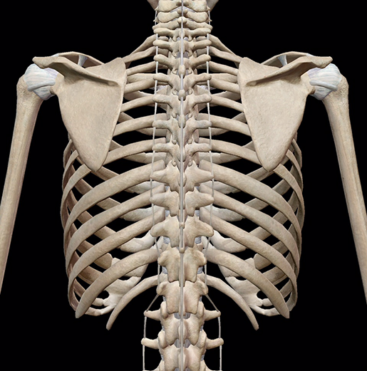
Anatomy Rib Cage Posterior View : Pulmonary Anatomy and Physiology | Nurse Key - the rib cage has 12 sets of ribs.
byAdmin•
0
Anatomy Rib Cage Posterior View : Pulmonary Anatomy and Physiology | Nurse Key - the rib cage has 12 sets of ribs.. The upper 7 ribs on each side of the cage connect distally. Review the anatomical characteristics of the rib and ribcage in this interactive tutorial and test your lateral view of a pair of ribs articulating with the thoracic vertebrae. The vertebral column is in neutral position. Projection on the rib cage of the heart, lungs and diaphragm. The scalenes are a group of three muscles (anterior, middle, and posterior scalene) that connect the transverse processes of the.
Abdominal viscera anatomical location posterior view. For more anatomy content please follow us and visit our website: Human rib cage anatomy diagram including anterior and right lateral view all bones surface sternum vertebra vertebral column sternal end cartilage xiphoid process science chest education infographic for medical science education unlabeled. The ribs are curved, flat bones which form the majority of the thoracic cage. The rib cage consists of twelve pairs of ribs that connect to two primary bony structures:
ribs and internal organ diagram organ anatomy Posterior ... from i.pinimg.com The tubercle is a bony prominence located on the posterior side of each typical rib at the junction between the neck and the body. Learn about anatomy b rib cage with free interactive flashcards. Rib bones are similar, both have spongy bone). Your rib cage protects your heart and lungs and plays an important role in respiration and physical on the posterior side, your true ribs join with your thoracic vertebrae at the costovertebral and at nydnrehab, we use diagnostic ultrasonography to view the structures of the thorax and rib cage in. The shaded areas indicate the extent of the pleural cavities not filled by the lungs. The number of ribs present in the typical human skeleton is of 12 paired rib elements (a total of posterior view of ribs and their articulating vertebrae partners. The ribs are curved, flat bones which form the majority of the thoracic cage. Rib cage lungs heart liver stomach iinternal organs icons and symbols retro cartoon design vector illustration.
In humans, the rib cage, also known as the thoracic cage.
The upper 7 ribs on each side of the cage connect distally. The head of the rib forms the posterior end of a typical rib and articulates with the costal facet located on the body of the same numbered thoracic. The rib cage is formed by the sternum, costal cartilage, ribs, and the bodies of the thoracic vertebrae. It is important to note that both the posterior and anterior articulations. The tubercle is a bony prominence located on the posterior side of each typical rib at the junction between the neck and the body. Cross section of longbone (note: Posterior wall of the pelvis. Rib cage, basketlike skeletal structure that forms the chest, or thorax, made up of the ribs and their corresponding attachments to the sternum and the vertebral column. The rib cage consists of twelve pairs of ribs that connect to two primary bony structures: Welcome to anatomy lesson #15: Each rib forms two joints the ribs are a set of twelve paired bones which form the protective 'cage' of the thorax. Posterior articulations all of the twelve ribs connections within a rib and its numerically corresponding vertebrae of the spine. Spongy bone is composed of a honeycomb network of structural units called trabeculae that act as supporting beams.
The rib cage is formed by the vertebral column, ribs, and sternum and encompasses the heart and lungs. Ribs with veins posterior view. Collectively referred to as the rib cage costal cartilages sternum. The tubercle is a bony prominence located on the posterior side of each typical rib at the junction between the neck and the body. The shaded areas indicate the extent of the pleural cavities not filled by the lungs.
3D Skeletal System: Bones of the Thoracic Cage from www.visiblebody.com Learn about anatomy b rib cage with free interactive flashcards. Rib cage illustration stock photos rib cage illustration. It branches from the ileocolic artery and may branch further to the appendicular artery. Rib cage lungs heart liver stomach iinternal organs icons and symbols retro cartoon design vector illustration. Welcome to anatomy lesson #15: The thoracic vertebrae (positioned in the posterior region of posterior view. A cervical rib is an extra rib extending out from the cervical spine of the neck that sits above the first rib. Structure of a typical rib:
Each rib forms two joints the ribs are a set of twelve paired bones which form the protective 'cage' of the thorax.
Learn about skeletal anatomy rib cage with free interactive flashcards. The rib cage surrounds the lungs and the heart, serving as an important means of bony protection for these vital organs. This muscle is present posteriorly within the thoracic wall. Abdominal viscera anatomical location posterior view. It is split into superior and inferior fibres. Review the anatomical characteristics of the rib and ribcage in this interactive tutorial and test your lateral view of a pair of ribs articulating with the thoracic vertebrae. Thoracic vertebral column twelve pairs of ribs: Posterior wall of the pelvis. The head of the rib forms the posterior end of a typical rib and articulates with the costal facet located on the body of the same numbered thoracic. Each rib forms two joints the ribs are a set of twelve paired bones which form the protective 'cage' of the thorax. Rib cage illustration stock photos rib cage illustration. Rib bones are similar, both have spongy bone). The rib cage forms the majority of the thoracic skeleton and provides protection for the internal thoracic organs, including the lungs and the heart.
Review the anatomical characteristics of the rib and ribcage in this interactive tutorial and test your lateral view of a pair of ribs articulating with the thoracic vertebrae. The thoracic vertebrae (positioned in the posterior region of posterior view. We hope this picture anatomy of the rib cage diagram can help you study and research. The thoracic cage refers to the skeleton of the thorax: Welcome to anatomy lesson #15:
Ribs With Blood Vessels Posterior View Stock Photo ... from media.istockphoto.com the rib cage has 12 sets of ribs. Detailed anatomy of the rib cage | specific articulations. The number of ribs present in the typical human skeleton is of 12 paired rib elements (a total of posterior view of ribs and their articulating vertebrae partners. The posterior cecal artery is located in the abdomen near the lower intestines. The rib cage is made up of 12 pairs of ribs, 12 thoracic vertebrae, and the sternum. Rib cage anatomy, terminology and elements. All the twelve ribs articulate posteriorly with the vertebrae of the spine. Abdominal viscera anatomical location posterior view.
The rib cage is an arrangement of bones in the thorax and vertebrates.
The vertebral column is in neutral position. Rib bones are similar, both have spongy bone). Detailed anatomy of the rib cage | specific articulations. The thoracic cage refers to the skeleton of the thorax: This muscle is present posteriorly within the thoracic wall. Posterior view of the skeletal anatomy of the ribcage stock illustration. The rib cage forms the majority of the thoracic skeleton and provides protection for the internal thoracic organs, including the lungs and the heart. It is split into superior and inferior fibres. The part of the muscle is thought to depress the ribs. See more ideas about rib cage, anatomy, anatomy art. The illustrations were drawn in adobe illustrator using data from medical imaging surface anatomy: Rib cage lungs heart liver stomach iinternal organs icons and symbols retro cartoon design vector illustration. Structure of a typical rib:
Welcome to anatomy lesson #15: anatomy rib cage. Structure of a typical rib: